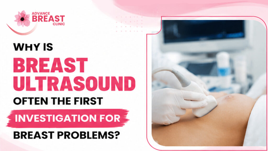
Why Is Breast Ultrasound Often the First Investigation for Breast Problems?
Breast ultrasound is often the first imaging investigation for certain breast problems because it is non-invasive, uses sound waves rather than ionizing radiation, and can provide real-time information about a breast lump or focal abnormality. It is particularly useful as initial imaging in younger women with a palpable breast mass and may also be useful during pregnancy or when further characterization of a breast finding is needed.
However, breast ultrasound is not automatically the first test for every woman and should not be considered a replacement for mammography in routine breast-cancer screening. The most appropriate investigation depends on age, symptoms, examination findings, breast density, previous imaging and individual breast-cancer risk.
Understanding that distinction helps explain why one woman may be sent directly for ultrasound while another may need mammography, ultrasound or both.
What Is a Breast Ultrasound?
A breast ultrasound is an imaging examination that uses high-frequency sound waves to create real-time pictures of structures inside the breast. It does not use X-rays or ionizing radiation. During the examination, a transducer is moved over the skin to create images of the underlying breast tissue.
Ultrasound can help doctors assess an area that feels different during examination or investigate something found on another breast-imaging test.
At Advance Breast Clinic, breast ultrasound forms part of a broader diagnostic pathway that can also include clinical examination, mammography, FNAC and biopsy when clinically indicated.
Why Is Breast Ultrasound Often Used as the First Investigation?
The key reason is not simply that ultrasound is “safe.” It is that ultrasound provides the type of information doctors need in several common clinical situations while avoiding unnecessary radiation exposure and allowing targeted real-time examination of the area of concern.
1. It is particularly useful for younger women with a breast lump
The American College of Radiology considers breast ultrasound usually appropriate initial imaging for a palpable breast mass in women younger than 30 years. Diagnostic mammography is generally not the preferred first investigation in this age group when evaluating a typical palpable mass.
Younger women tend to have more fibroglandular breast tissue, and common benign causes of breast lumps, such as fibroadenomas and cysts often occur in this age group.
Ultrasound can provide useful information about the lump without immediately exposing a young patient to X-rays.
2. It can distinguish fluid-filled from solid findings
One of ultrasound’s practical strengths is its ability to help characterize a palpable or imaging-detected lesion.
For example, ultrasound may help determine whether a finding behaves like:
- A simple fluid-filled cyst
- A solid breast mass
- A complex cystic and solid lesion
- Normal breast tissue
- Another focal abnormality requiring further assessment
RadiologyInfo, an educational resource from the American College of Radiology and Radiological Society of North America, notes that ultrasound can help determine whether an area of concern is normal tissue, a benign cyst or another lesion that may require further investigation.
Importantly, “solid” does not automatically mean cancer. Many solid breast lumps are benign.
3. It provides real-time imaging
Unlike a static image, ultrasound allows the operator to examine a specific area dynamically.
A clinician or radiologist can focus directly on the spot where the patient feels a lump or where examination identified an abnormality.
This real-time capability also makes ultrasound useful for guiding a needle accurately into a lesion when tissue sampling is required.
4. It does not use ionizing radiation
Standard diagnostic breast ultrasound uses sound waves and has no ionizing radiation exposure. The ACR lists breast ultrasound with a relative radiation level of 0 mSv in its appropriateness criteria.
This makes ultrasound especially useful when radiation avoidance is desirable, although the decision about which imaging study is best should still be based on clinical appropriateness rather than radiation alone.
5. It is useful during pregnancy
When a pregnant woman develops a focal breast problem or palpable lump, ultrasound frequently plays an important role in initial imaging. The ACR rates breast ultrasound as usually appropriate initial imaging for pregnant women with focal breast pain or a palpable mass, including women younger than 30.
This does not mean mammography can never be used during pregnancy. Depending on age and clinical suspicion, additional imaging may still be appropriate.
Who Commonly Has Breast Ultrasound as the First Imaging Test?
The answer depends mainly on age, symptoms and clinical context.
| Clinical situation | Typical role of ultrasound |
| Woman under 30 with a new palpable lump | Often the first imaging test |
| Woman aged 30–39 with a palpable lump | Ultrasound and/or diagnostic mammography may be appropriate |
| Woman 40+ with a new palpable lump | Diagnostic mammography/DBT usually has an important initial role; ultrasound is often complementary |
| Pregnant woman with focal lump or pain | Ultrasound often plays a first-line role |
| Abnormality seen on mammography | Targeted ultrasound may further characterize it |
| Dense breasts | Ultrasound may sometimes be supplemental, depending on risk |
| Suspicious ultrasound-visible mass | Ultrasound may guide tissue sampling |
These distinctions are reflected in ACR Appropriateness Criteria for palpable breast masses, pregnancy and breast density.
The main takeaway is simple:
The best first investigation is selected for the patient—not just for the symptom.
What Can Breast Ultrasound Reveal About a Lump?
A breast ultrasound can provide several useful pieces of information about a localized breast finding.
It can help assess:
- Whether a lesion is fluid-filled or solid
- Its size
- Its shape
- Its orientation
- Its margins
- Its relationship with surrounding tissue
- Features that appear more reassuring or more suspicious
- Nearby lymph nodes when clinically relevant
The imaging findings are interpreted in context. Ultrasound cannot independently provide a tissue diagnosis for every lesion.
A clearly benign-appearing cyst, for example, may need very different management from a suspicious irregular solid mass.
That is why imaging classification guides the next step rather than replacing clinical judgment or biopsy when biopsy is indicated.
Breast Ultrasound vs Mammography: Which Is Better?
Neither test is universally “better.” They answer different clinical questions and are often complementary.
Breast ultrasound
Breast ultrasound:
- Uses sound waves
- Has no ionizing radiation
- Provides real-time imaging
- Is particularly useful for localized symptoms and palpable lumps
- Can differentiate fluid-filled and solid findings
- Can guide biopsy procedures
Mammography
Mammography:
- Uses low-dose X-rays
- Provides a wider overview of breast tissue
- Can detect masses and architectural distortion
- Is particularly important for identifying microcalcifications
- Is the primary established imaging method for routine population breast-cancer screening in appropriate age groups
Ultrasound does not reliably display all findings that mammography can detect. Certain early cancers may present mainly as calcifications, which may be visible on mammography but not on ultrasound.
For women aged 40–74 at average breast-cancer risk, the USPSTF recommends screening mammography every two years. That recommendation concerns screening women without symptoms, which is different from investigating a new lump.
Advance Breast Clinic already provides patient information explaining the distinction between breast screening and diagnostic testing. Screening aims to detect disease before symptoms appear, whereas diagnostic imaging investigates an existing symptom or abnormal finding.
For readers who want to understand mammography in more detail, see the clinic’s guide to mammography in Nepal.
What About Breast Ultrasound for Dense Breasts?
Dense breast tissue can make mammographic interpretation more challenging because dense fibroglandular tissue can obscure some abnormalities.
Ultrasound may sometimes be considered as a supplemental examination in women with dense breasts, but breast density alone does not mean every woman needs ultrasound.
The ACR currently rates ultrasound as potentially appropriate supplemental screening in some women with heterogeneously or extremely dense breasts, depending on overall breast-cancer risk. MRI may be preferred in some higher-risk situations.
The USPSTF, meanwhile, states that evidence is currently insufficient to determine the balance of benefits and harms of universally adding ultrasound or MRI after a negative mammogram in women with dense breasts.
Therefore:
Dense breasts are a reason for individualized risk assessment not an automatic prescription for ultrasound.
Is Breast Ultrasound Safe?
Standard diagnostic ultrasound does not expose the patient to ionizing radiation, and RadiologyInfo reports no known harmful effects from standard diagnostic breast ultrasound.
The examination is non-invasive.
During the scan:
- You normally lie on an examination table.
- Gel is applied to the breast.
- A handheld ultrasound transducer is moved across the area.
- Real-time images appear on the monitor.
- Particular attention is given to the area of concern.
Advance Breast Clinic’s own breast-ultrasound information also describes ultrasound as a non-invasive examination using a transducer placed over the breast.
What Happens Before the Ultrasound?
Imaging is only one part of breast assessment.
A patient with a breast complaint may first undergo a clinical breast examination, during which the clinician asks about symptoms and examines the breasts and underarm regions. Factors such as age, duration of the lump, breast pain, nipple discharge, menstrual history, family history and previous imaging can influence which investigation is chosen.
This is why ordering the same test for every breast symptom is not ideal.
A focused clinical breast examination can help determine whether ultrasound, mammography or another investigation is the appropriate next step.
What Happens If Breast Ultrasound Finds a Lump?
Not every finding leads to biopsy.
The next step depends on how the abnormality appears and whether the imaging findings match the clinical concern.
Possible pathways include:
Clearly benign finding
A simple cyst or another confidently benign finding may require no invasive procedure, although management depends on symptoms and the overall clinical picture.
Probably benign finding
Some findings may be considered probably benign and monitored with short-term follow-up imaging rather than immediate biopsy. ACR patient guidance notes that BI-RADS 3 findings commonly enter an imaging follow-up pathway.
Suspicious finding
If ultrasound identifies a suspicious abnormality, additional imaging or tissue sampling may be recommended.
For women younger than 30 with a suspicious ultrasound finding, the ACR considers diagnostic mammography/tomosynthesis and image-guided core biopsy appropriate next steps.
This illustrates an important point:
Ultrasound may start the investigation, but it does not necessarily finish it.
A new breast lump should not be evaluated by choosing an imaging test based only on age or online information. A specialist breast assessment can combine your symptoms, clinical examination, previous imaging and risk factors to decide whether ultrasound, mammography or tissue sampling is appropriate.
How Can Ultrasound Guide FNAC or Breast Biopsy?
One major advantage of ultrasound is that it can provide continuous real-time visualization during needle procedures.
If a suspicious lesion can be seen clearly on ultrasound, the doctor can use the ultrasound image to guide a needle toward the correct area.
Two terms patients may encounter are:
FNAC
Fine-needle aspiration cytology uses a thin needle to collect cells from a mass for microscopic examination. Advance Breast Clinic describes FNAC as a relatively minimally invasive method of obtaining cellular material for diagnostic assessment.
Core needle biopsy
A core biopsy obtains small pieces of tissue rather than only individual cells.
For many suspicious solid breast lesions, tissue architecture can provide information that cytology alone cannot.
Which procedure is appropriate depends on the lesion and clinical question and should be selected by the treating team.
Ultrasound-guided sampling
During an ultrasound-guided FNAC or biopsy, ultrasound helps the clinician see the lesion and guide the sampling needle to the intended location.
RadiologyInfo likewise notes that ultrasound guidance is commonly used for breast biopsy because the technique provides real-time visualization and does not use ionizing radiation.
These procedures are generally minimally invasive, but they are still medical procedures and can carry risks such as bleeding or infection.
Can Breast Ultrasound Detect Breast Cancer?
Breast ultrasound can identify suspicious abnormalities, but ultrasound alone cannot reliably detect or exclude every breast cancer.
This is one of the most important limitations to understand.
Ultrasound can help characterize many masses and may demonstrate suspicious features. However:
- Some cancers may not be visible on ultrasound.
- Some early cancers appear mainly as mammographic calcifications.
- A suspicious ultrasound finding may still require biopsy for definitive diagnosis.
- A normal ultrasound does not automatically override a clinically concerning breast change.
RadiologyInfo specifically states that ultrasound does not replace mammography and that many calcifications seen on mammography cannot be seen with ultrasound.
Likewise, ACR guidance emphasizes that a new palpable lump may still require further clinical follow-up even when initial imaging appears normal.
Therefore, patients should avoid interpreting “normal ultrasound” as meaning that every possible breast disease has been excluded.
Is Breast Ultrasound a Breast-Cancer Screening Test?
Breast ultrasound can have a supplemental screening role in selected women, but it is not the standard universal primary screening test for average-risk women.
For routine screening, mammography remains the better-established population screening method. The USPSTF recommends biennial mammography for average-risk women aged 40–74.
Women with higher inherited risk, previous breast cancer or other major risk factors may require a different imaging strategy involving MRI, mammography or other testing under specialist guidance.
This is why the words screening and investigation should not be used interchangeably.
- Screening: testing someone without symptoms.
- Diagnostic investigation: evaluating a current symptom, lump or abnormal finding.
Advance Breast Clinic’s existing breast screening guide explains this same distinction.
Why a Breast Lump Needs More Than an Imaging Test
The safest breast-lump pathway is not:
Feel lump → automatically get ultrasound → assume diagnosis
A better clinical framework is:
History → clinical examination → age-appropriate imaging → correlate findings → biopsy when indicated
This approach matters because not every breast lump is cancer and not every cancer has a classic appearance on one imaging modality.
Breast lumps can include cysts, fibroadenomas and other benign conditions as well as malignant disease. Advance Breast Clinic also addresses these different presentations in its information on common breast-related problems.
The goal of breast assessment is therefore not to perform as many tests as possible.
It is to choose the right investigation for the right clinical problem.
When Should You See a Breast Specialist?
Arrange medical evaluation if you notice a new or persistent breast change such as:
- A new lump
- A lump in the underarm
- Persistent focal breast change
- Nipple inversion
- Spontaneous or bloody nipple discharge
- Skin dimpling or thickening
- Unexplained change in breast shape
- Persistent redness or swelling
- An abnormal mammogram or ultrasound report
These findings do not automatically mean breast cancer, but they warrant proper assessment rather than observation without a plan. Advance Breast Clinic’s first-visit guidance similarly recommends assessment when a lump, nipple discharge, skin dimpling or nipple change is noticed.
For patients in Kathmandu Valley, Advance Breast Clinic is located in Jawalakhel, Lalitpur and lists consultation and breast diagnostic services including clinical examination, ultrasound, mammography and biopsy pathways.
Key Takeaways
Breast ultrasound is often chosen first because it is:
- Non-invasive
- Free from ionizing radiation
- Effective for targeted real-time assessment
- Particularly useful for breast lumps in younger women
- Helpful in distinguishing cystic from solid findings
- Useful during pregnancy
- Able to guide FNAC and biopsy
But breast ultrasound is not the first test in every patient and does not replace mammography for routine breast-cancer screening. Imaging should be selected according to age, symptoms, examination findings, breast-cancer risk and prior results.
The most useful question is therefore not:
“Is ultrasound better than mammography?”
It is:
“Which investigation is most appropriate for my age, symptoms and breast findings?”
FAQs
Why is breast ultrasound usually preferred for younger women with a breast lump?
For women younger than 30 with a palpable breast mass, ACR guidance considers breast ultrasound usually appropriate as the initial imaging examination. It provides targeted assessment without ionizing radiation and can help characterize whether a lump is fluid-filled or solid. Further imaging or biopsy may still be necessary if suspicious features are found.
Is breast ultrasound safer than mammography?
Breast ultrasound does not use ionizing radiation, whereas mammography uses a low dose of X-rays. However, “safer” does not mean “better” for every situation. Mammography can detect findings such as microcalcifications that ultrasound may miss, making the two tests complementary rather than interchangeable.
Can breast ultrasound tell whether a lump is cancer?
Ultrasound can show whether a lesion has features that appear benign, probably benign or suspicious, but imaging alone cannot definitively diagnose every breast cancer. If a lesion is suspicious, tissue sampling may be necessary to determine its nature.
Can breast ultrasound distinguish a cyst from a solid lump?
Yes. Distinguishing fluid-filled cystic findings from solid lesions is one of the useful applications of breast ultrasound. However, solid does not automatically mean malignant, and complex findings may need additional imaging or biopsy.
Is ultrasound enough if I have dense breasts?
Not necessarily. Ultrasound can sometimes supplement mammography in women with dense breasts, but the need depends on overall breast-cancer risk. Mammography remains important, and some higher-risk women may benefit more from MRI or another supplemental strategy.
Is breast ultrasound used during pregnancy?
Yes. Ultrasound commonly plays an important initial role when a pregnant woman develops a focal breast problem or palpable mass because it does not use ionizing radiation. Depending on age and clinical concern, additional breast imaging may still be appropriate.
Does a normal breast ultrasound mean the lump is definitely harmless?
No. Imaging findings must match the clinical examination. ACR guidance advises continued medical follow-up for a new palpable mass even when imaging appears normal if the clinical concern remains.
Can biopsy be done using breast-ultrasound guidance?
Yes. When a lesion is visible on ultrasound and tissue sampling is required, real-time ultrasound can guide the needle into the target area for FNAC or core biopsy.