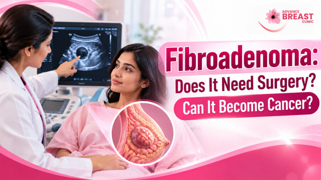
Fibroadenoma: Does It Need Surgery? Can It Become Cancer?
Being told that a breast lump is probably a fibroadenoma can bring two immediate questions: Will I need surgery? and Could it become breast cancer?
The reassuring answer is that a fibroadenoma is a benign, noncancerous breast tumour, and many fibroadenomas do not need to be surgically removed. The National Cancer Institute describes fibroadenoma as a benign tumour made from glandular and connective breast tissue, most commonly found in younger women.
Whether a particular fibroadenoma should simply be observed, investigated further or removed depends on more than its name. Doctors consider how confidently the diagnosis has been established, whether the lump is growing, whether it causes symptoms, its size, imaging and biopsy findings, and the patient’s preferences.
The important first step is therefore not automatically choosing surgery. It is making sure that the breast lump has been appropriately evaluated.
What Is a Fibroadenoma?
A fibroadenoma is a solid benign breast mass made up of fibrous or connective tissue and glandular breast tissue.
It often feels smooth, firm or rubbery and can move beneath the skin when pressed. Many are painless. Fibroadenomas occur most commonly in younger women, although they can occur at other ages as well.
Fibroadenomas can behave differently over time. Some remain almost unchanged for years. Others may become smaller, and some eventually disappear. Hormonal changes can also influence them. They may enlarge during pregnancy and often become smaller after menopause.
Having a lump that feels movable or smooth does not, however, prove that it is a fibroadenoma. Different breast conditions can overlap in how they feel.
If you have found a new breast lump, the goal is to establish what the lump actually represents rather than trying to diagnose it by touch alone. Advance Breast Clinic’s guide to the different causes of breast lumps explains why clinical assessment and appropriate imaging matter.
How Is a Fibroadenoma Diagnosed?
Evaluation generally begins with your medical history and a clinical breast examination.
The clinician may ask:
- how long the lump has been present
- whether it has changed in size
- whether it is painful
- whether you are pregnant or breastfeeding
- whether you have previously had fibroadenomas
- whether you have relevant family or personal breast history and
- whether you have noticed nipple, skin or breast-shape changes.
Breast imaging
Imaging helps determine the size, shape and characteristics of a breast mass.
Breast ultrasound is particularly useful for evaluating many lumps in younger patients because it can characterize a solid mass and distinguish it from a fluid-filled cyst. The appropriate test nevertheless depends on the person’s age, symptoms, risk factors and clinical findings. Mammography may also be appropriate in some situations.
For a more detailed explanation, see the clinic’s guide to breast ultrasound and when it is used.
If your report contains a BI-RADS assessment, the clinic’s BI-RADS guide explains how those imaging categories are used.
Is a biopsy always necessary?
No.
Some breast masses may have sufficiently typical features that the clinician and radiologist can recommend observation rather than immediate tissue sampling. In other situations, a biopsy is needed to establish the diagnosis with greater certainty.
Core needle biopsy obtains small samples of tissue that a pathologist examines under a microscope. This can help distinguish fibroadenoma from other fibroepithelial lesions, including a phyllodes tumour.
A particularly important concept is concordance. This means the clinical picture, imaging findings and biopsy result make sense together.
For example, if imaging looks suspicious but the biopsy result seems unexpectedly benign, doctors may need to investigate further rather than simply accepting the word “fibroadenoma.”
You can read more about tissue sampling in the clinic’s guide to breast biopsy in Nepal.
Does Every Fibroadenoma Need Surgery?
No. Many fibroadenomas can safely remain in the breast when the diagnosis is secure and there is no other reason for removal.
This is an important change from the assumption that every solid breast lump must be surgically removed.
The 2025 American Society of Breast Surgeons and Society of Breast Imaging guideline reached strong consensus that a core-biopsy-proven fibroadenoma without atypia, when the pathology and imaging are concordant, generally needs excision only when factors such as symptoms, substantive growth, size or patient preference justify it.
When observation may be reasonable
Observation may be appropriate when:
- the diagnosis is considered reliable
- imaging and pathology findings agree
- the lesion is not causing significant symptoms
- it is not showing concerning growth or other change and
- there is no clinical concern for a different type of tumour.
Observation does not mean ignoring a breast lump.
It means a clinician has assessed the available evidence and concluded that immediate removal is unlikely to provide enough benefit to justify surgery.
Surgery itself has trade-offs. Removing a fibroadenoma can leave a scar, alter breast contour or remove some surrounding normal breast tissue. Mayo Clinic therefore notes that the cosmetic effects of surgery should be considered when deciding whether removal is necessary.
When fibroadenoma removal may be considered
Removal may become reasonable when a fibroadenoma
- causes troublesome discomfort or other symptoms
- becomes large enough to distort breast shape
- demonstrates substantial or rapid growth
- has imaging or biopsy findings that do not fit comfortably together
- cannot confidently be distinguished from another fibroepithelial lesion or
- causes enough ongoing concern that, after an informed discussion of benefits and limitations, the patient prefers excision.
The exact decision is individualized.
There is no universal rule that every fibroadenoma above one particular diameter must automatically be removed. Size is one consideration within the complete clinical picture.
Stable confirmed fibroadenoma versus changing lesion
| Situation | Typical management question |
|---|---|
| Confirmed fibroadenoma with concordant imaging/pathology and no significant symptoms | Is surgery providing any meaningful benefit? |
| Significant symptoms | Would removal improve quality of life enough to justify surgery? |
| Substantial growth | Does the lesion still behave consistently with fibroadenoma, or should it be reassessed? |
| Uncertain or discordant diagnosis | Is additional tissue assessment or excision needed to establish the correct diagnosis? |
| Concern for phyllodes tumour | Is complete surgical excision appropriate? |
| Patient strongly prefers removal after counseling | What are the cosmetic and procedural trade-offs? |
This is why two people who both have “fibroadenoma” written on their reports may appropriately receive different recommendations.
Can Fibroadenoma Become Cancer?
A typical fibroadenoma is not breast cancer and is not generally considered a lesion that routinely transforms into cancer.
The National Cancer Institute states that most fibroadenomas do not increase breast-cancer risk. The American Cancer Society similarly notes that simple fibroadenomas do not appear to increase risk.
The subject becomes more nuanced when pathology reports mention a complex fibroadenoma or other proliferative changes.
Older observational research found higher subsequent breast-cancer rates in some women whose excised fibroadenomas had complex features or who also had proliferative breast disease. Later research has suggested that associated breast-tissue characteristics and other risk factors are important when interpreting this association.
For an individual patient, this means the word fibroadenoma should not be interpreted in isolation.
Your overall breast-cancer risk may depend on factors such as:
- the exact pathology
- whether atypical or proliferative changes are present elsewhere
- age
- family history
- genetic risk where relevant and
- other established breast-cancer risk factors.
A simple fibroadenoma itself should not be treated as though it were an early breast cancer.
Fibroadenoma is not the same as a phyllodes tumour
This distinction matters.
Fibroadenomas and phyllodes tumours are both fibroepithelial breast lesions and can sometimes resemble one another. Most phyllodes tumours are benign, but some can be borderline or malignant.
The 2025 ASBrS/SBI guideline therefore recommends surgical excisional biopsy for lesions where there is concern for a phyllodes tumour rather than managing them automatically as ordinary fibroadenomas.
This is one reason a rapidly changing or diagnostically uncertain “fibroadenoma” deserves reassessment rather than reassurance based only on the original label.
What If a Fibroadenoma Starts Growing?
Growth does not automatically mean cancer.
Fibroadenomas can enlarge, particularly in hormonally active periods, including pregnancy. Younger patients may also experience changing lesions over time.
Nevertheless, meaningful growth deserves medical review.
The important questions are:
Has the lesion changed substantially?
Do its new imaging characteristics still look typical?
Was the original diagnosis made by imaging alone or confirmed by tissue?
Do pathology and imaging remain concordant?
Could the lesion represent another type of fibroepithelial tumour?
Current breast-surgery guidance includes substantive growth among the reasons clinicians may consider excision of an otherwise benign fibroadenoma.
Therefore, if a lump previously diagnosed as fibroadenoma becomes noticeably larger, do not assume either that it has become cancer or that growth can safely be ignored. Arrange reassessment.
Fibroadenoma During Pregnancy
Fibroadenomas can enlarge during pregnancy because breast tissue responds to hormonal changes. This enlargement does not by itself establish malignancy.
The 2025 benign fibroepithelial-lesion guideline recommends managing fibroadenomas diagnosed during pregnancy or before a planned pregnancy similarly to typical fibroadenomas, while persistent rapid growth warrants reevaluation and may justify repeat biopsy.
Because breast tissue changes considerably during pregnancy and lactation, any new or changing breast mass should be discussed with an appropriate clinician rather than attributed to pregnancy without assessment.
Fibroadenoma in Teenagers and Young Women
Fibroadenomas are particularly common in adolescents and young adults.
Conservative management is often possible for a stable lesion that has been appropriately assessed. Surgery may be considered for symptomatic, large or rapidly enlarging masses, but clinicians also consider the potential effect of an operation on the developing breast.
A young age should therefore reduce unnecessary alarm, but it should not substitute for assessment of a persistent or changing mass.
What Happens If a Fibroadenoma Is Removed?
The conventional surgical approach is excision of the mass.
The 2025 ASBrS/SBI guideline recommends complete removal without transecting the lesion when surgery is performed and advises surgeons to consider aesthetics, sensation and incision placement.
Important questions to discuss before surgery include:
- Why is removal being recommended?
- Has the diagnosis been confirmed?
- What incision is planned?
- Where is the scar likely to be?
- Could removal alter breast shape?
- Will the entire lesion be sent for pathology?
- What recovery should I expect?
Removal of one fibroadenoma also does not guarantee that another fibroadenoma will never appear. New fibroadenomas can develop separately over time.
When Should You See a Breast Specialist?
Arrange assessment if you notice:
- a new breast lump
- a known lump that has clearly grown or changed
- persistent focal breast changes
- unexplained nipple discharge
- a new nipple change
- skin dimpling or distortion
- unexplained change in breast shape
- a lump in the armpit or
- an imaging or biopsy result that you do not understand.
These symptoms do not automatically mean breast cancer. They mean the finding deserves an appropriate clinical explanation.
Fibroadenoma Assessment in Nepal
If you have found a breast lump, have been told that an ultrasound suggests fibroadenoma, or are unsure whether surgery is necessary, the useful next step is a breast-focused clinical assessment rather than deciding on treatment from an internet description alone.
Advance Breast Clinic in Jawalakhel, Lalitpur lists clinical breast examination, ultrasound, mammography, FNAC/biopsy and breast-surgery assessment among its breast-care services.
You can learn about clinical breast consultation at Advance Breast Clinic or review the verified professional profile of Dr. Banira Karki.
The bottom line
A fibroadenoma is usually benign and does not automatically need surgery.
When the diagnosis is well established and the lesion is stable and not troublesome, observation may be appropriate. Surgery becomes more relevant when a fibroadenoma is symptomatic, substantially growing, large enough to create problems, diagnostically uncertain or when removal is the informed preference of the patient.
A typical fibroadenoma is not breast cancer. The most useful question is therefore not simply “Should every fibroadenoma be removed?” but:
How certain is my diagnosis, has the lump changed, and what benefit would surgery provide in my particular case?
FAQs
Can fibroadenoma disappear without treatment?
Yes. Some remain stable, some shrink, and some can eventually disappear. NCI and Mayo both note that fibroadenomas may decrease or resolve without removal.
Does fibroadenoma always hurt?
No. Many are painless, although some people experience tenderness or discomfort.
Can I have more than one fibroadenoma?
Yes. A person can have multiple fibroadenomas and they may occur in one or both breasts.
Will surgery prevent fibroadenoma from returning?
Removing one mass removes that particular lesion, but a new fibroadenoma can develop elsewhere later.
Does a growing fibroadenoma mean cancer?
Not necessarily. Fibroadenomas can grow for benign reasons, but substantive or persistent growth warrants reassessment because diagnosis and management may need to be reconsidered.
Medical disclaimer: This article provides general educational information and does not diagnose a breast lump or replace individual medical advice. New, persistent or changing breast findings should be assessed by an appropriate healthcare professional.